Chapter 1
Structure and Function of Exercising Muscle
When the heart beats, when partially digested food moves through the intestines, and when the body moves in any way, muscle is involved. These many and varied functions of the muscular system are performed by three distinct types of muscle : smooth muscle, cardiac muscle, and skeletal muscle.
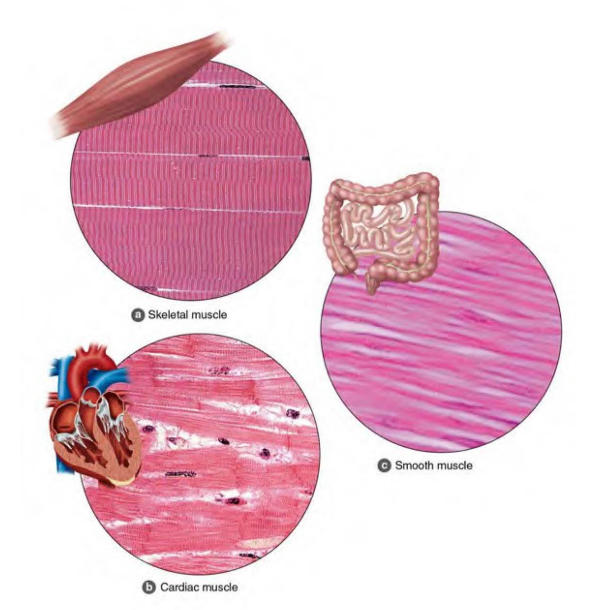
Anatomy of Skeletal Muscle
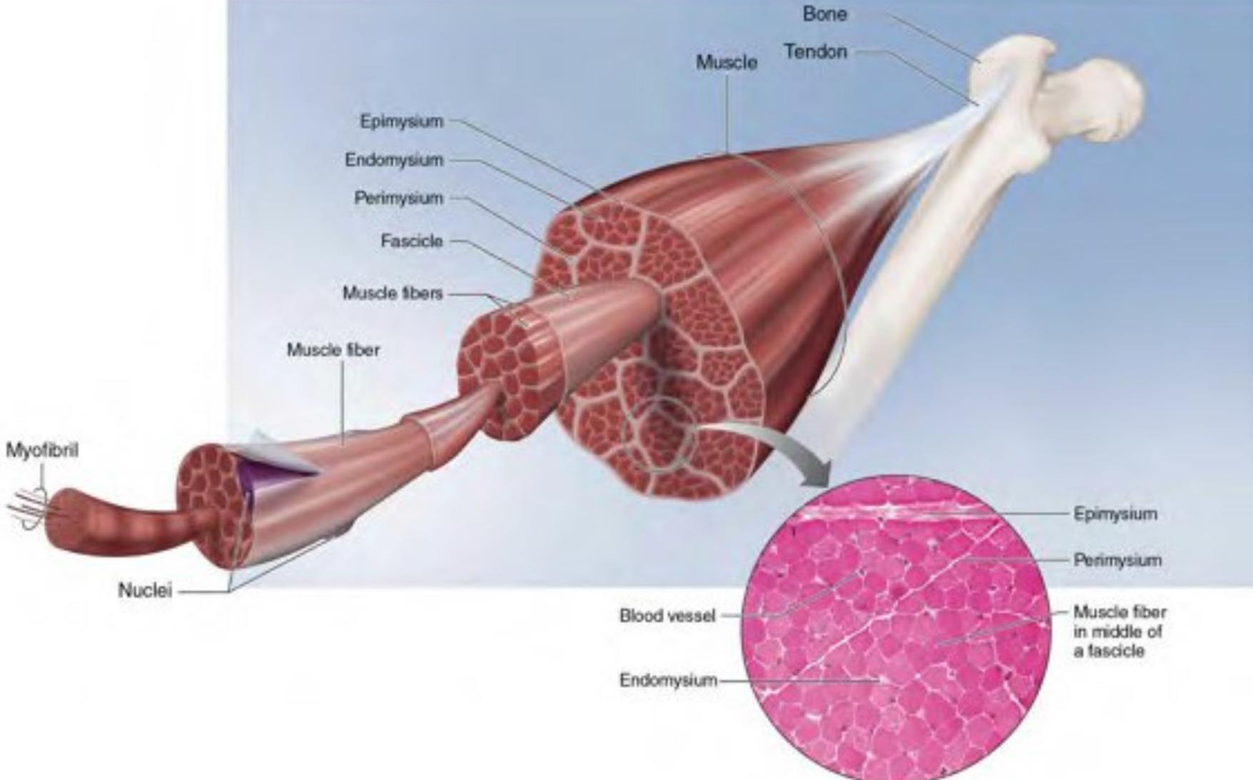
An individual muscle cell is called a muscle fiber.
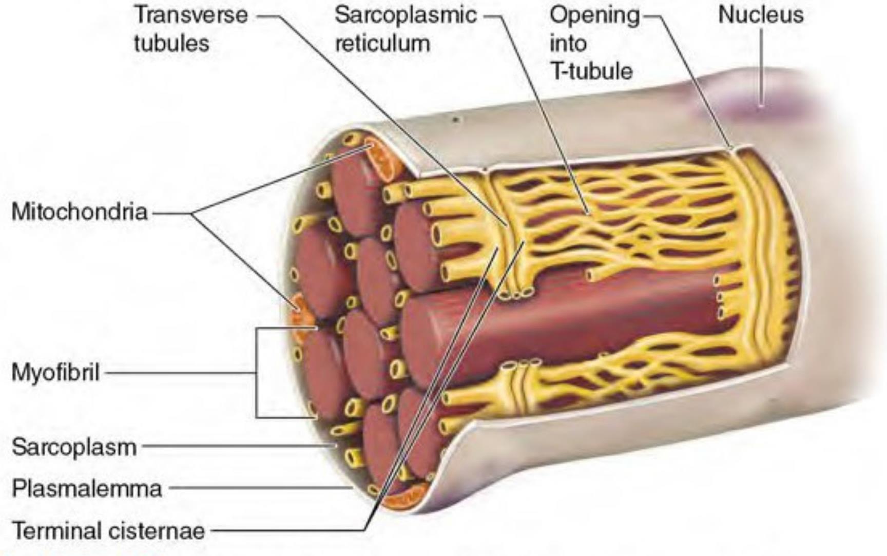
Muscle fibers have a cell membrane and the same organelles mitochondria, lysosomes, and so on- -as other cell types but are uniquely multinucleated. .
A muscle fiber is enclosed by a plasma membranecalled the plasmalemma.
The cytoplasm of a muscle fiber is called thesarcoplasm.
The extensive tubule network found in the sarcoplasmincludes T-tubules, which allow communication and transport of substances throughout the muscle fiber, andthe SR, which stores calcium.
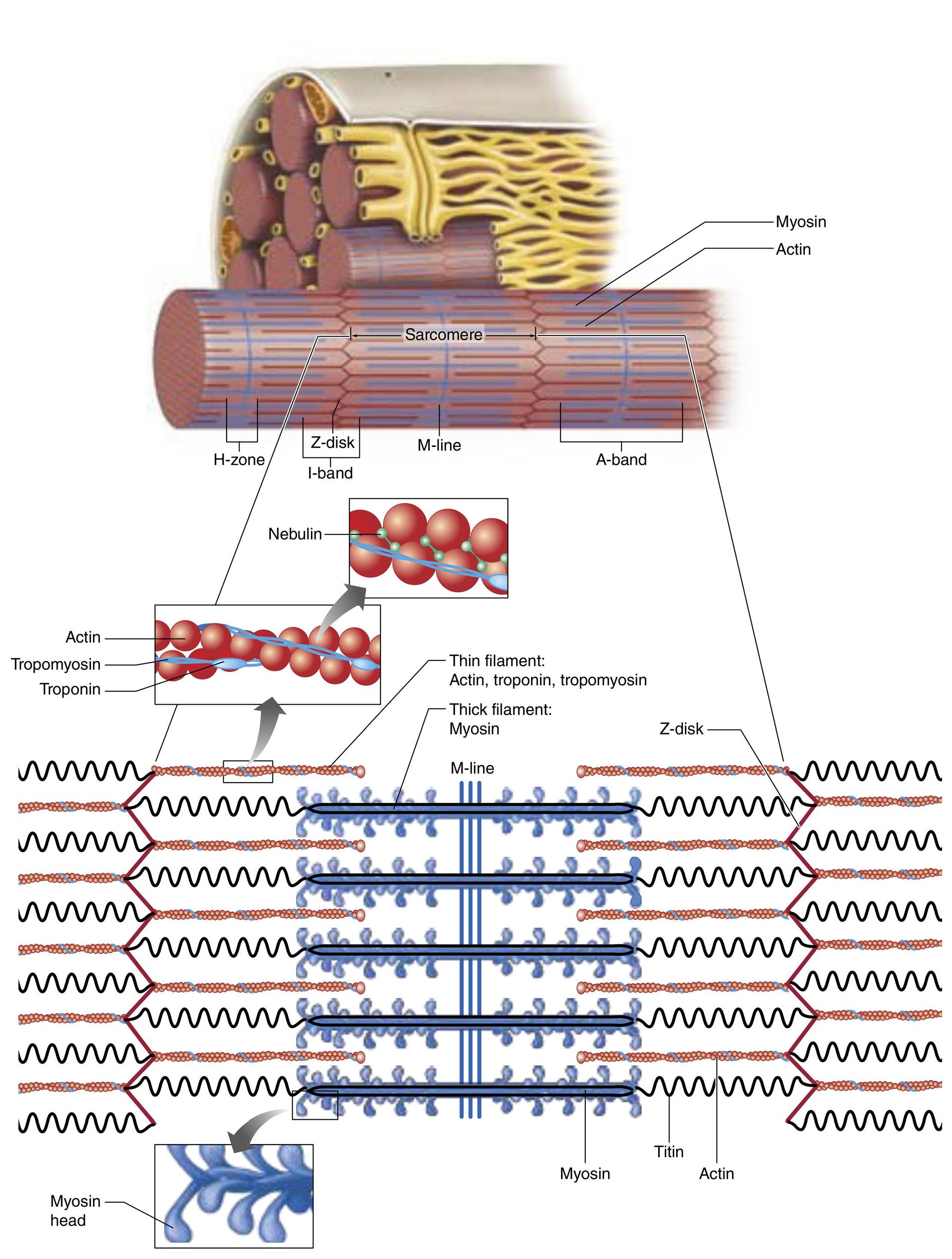
The sarcomere is the smallest functional unit of amuscle.
ThickFilaments Thin Filaments
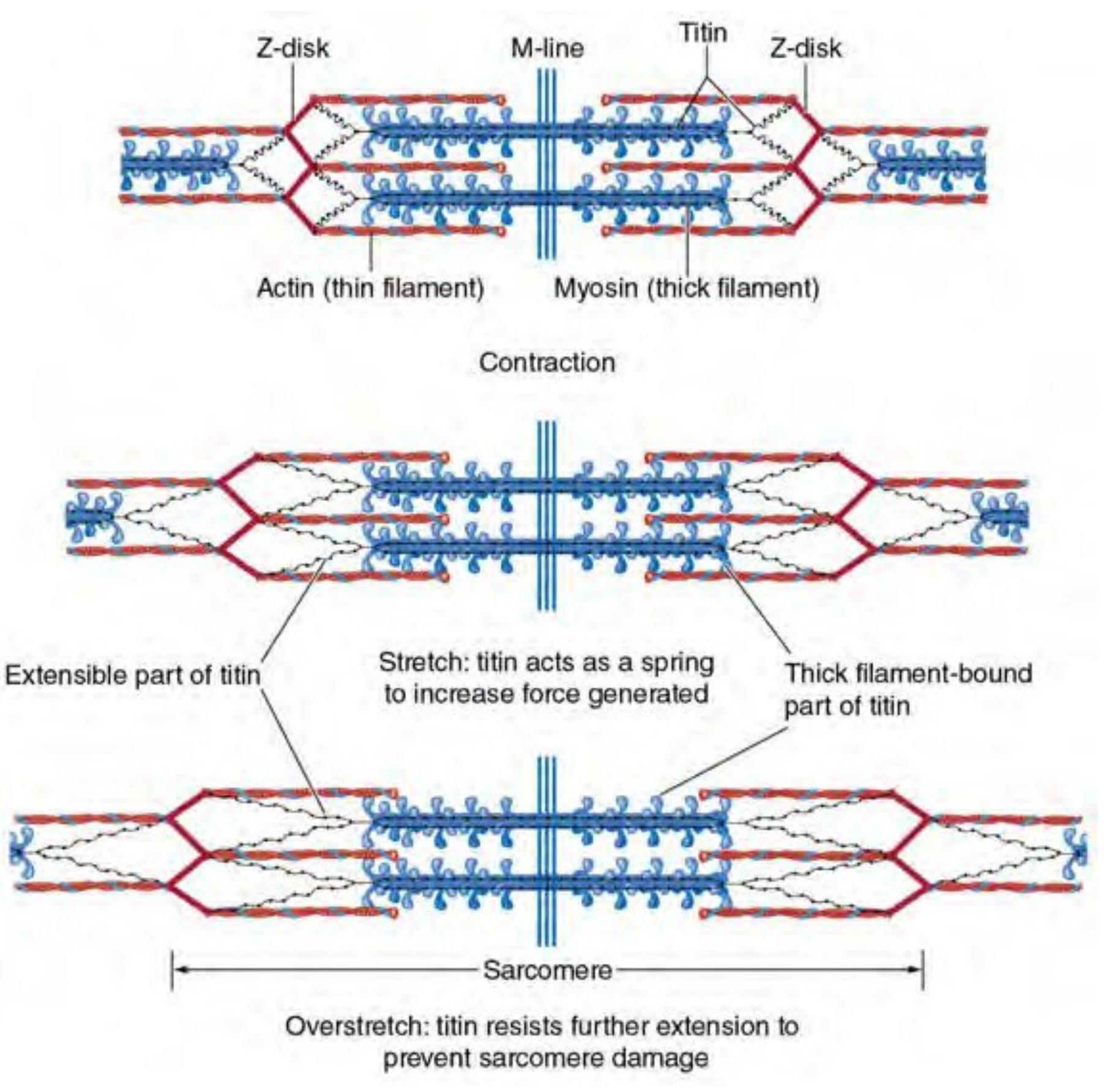
The mechanism through which the molecule titin acts during muscle contraction.Titin acts as a spring element to increase the force generated and resistsoverstretch to prevent sarcomere damage.
Myofibrils are composed of sarcomeres, thebasic contractile units of a muscle.
A sarcomere is composed of two different-sized filaments, thick and thin filaments, which are responsible for musclecontraction.
Myosin, the primary protein of the thickfilament, is composed of two protein strands, each folded into a globular headat one end.
The thin filament is composed of actin,tropomyosin, and troponin. One end of each thin filament is attached to aZ-disk.
Muscle Fiber Contraction
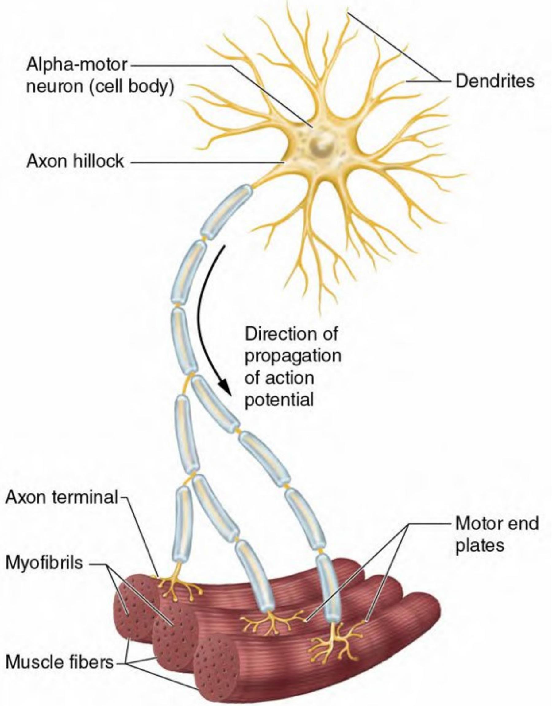
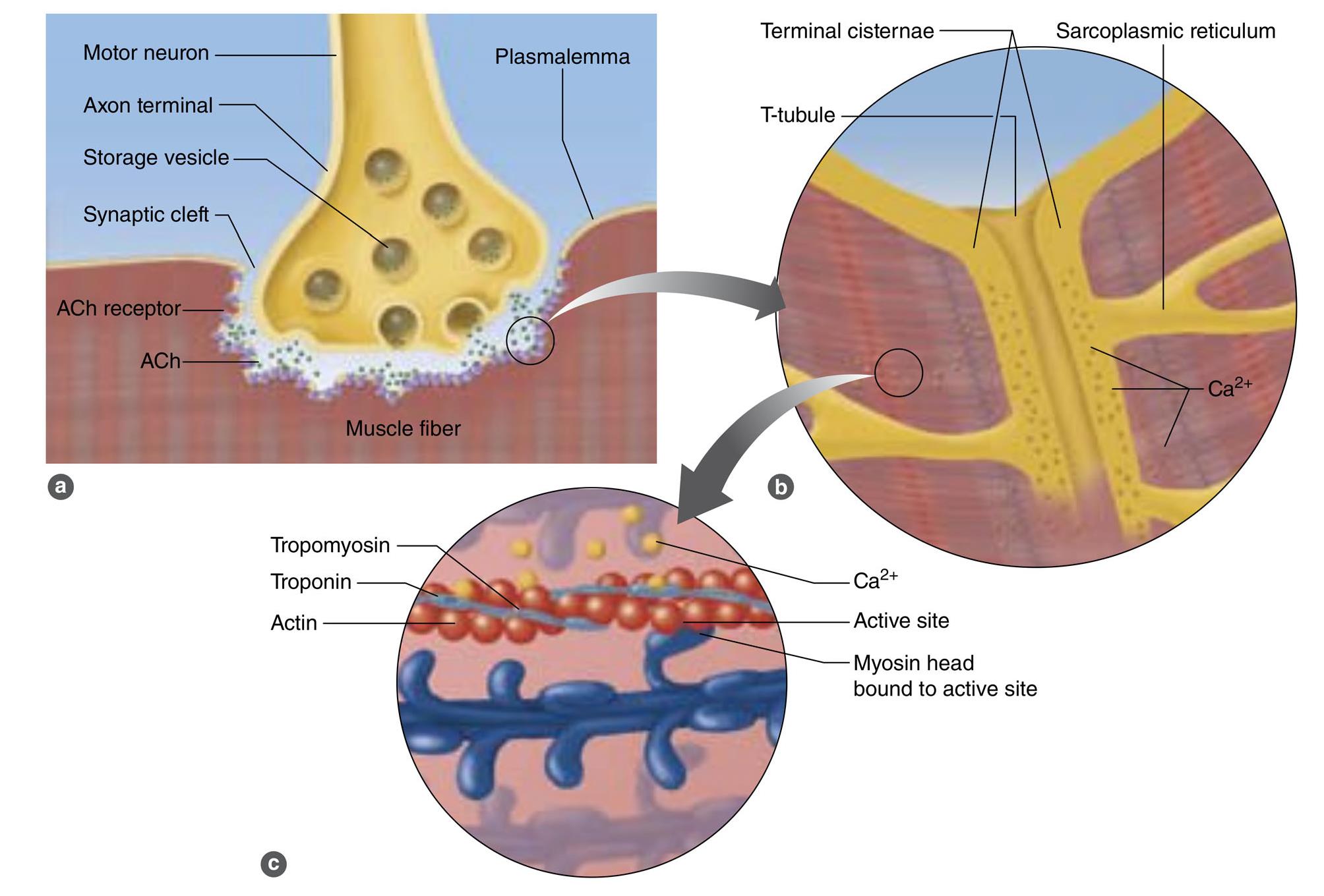
The sequence of events leading to muscleaction, known as excitation-contraction coupling.
(a) In response to an action potential, amotor neuron releases acetylcholine (ACh), which crosses the synaptic cleft andbinds to receptors on the plasmalemma. If enough ACh binds, an action potentialis generated in the muscle fiber.
(b) The action potential triggers the releaseof calcium ions (Ca2+) from the terminal cisternae of thesarcoplasmic reticulum into the sarcoplasm.
(c) The Ca2+ binds to troponin onthe actin filament, and the troponin pulls tropomyosin off the active sites,allowing myosin heads to attach to the actin filament.
The Sliding Filament Theory: How Muscles Create Movement
When muscle contracts, muscle fibers shorten. How do they shorten? The explanation for this phenomenon is termed the sliding filament theory. When the myosincross-bridges are activated, they bind with actin, resulting in aconformational change in the cross-bridge, which causes the myosin head to tiltand to drag the thin filament toward the center of the sarcomere .This tilting of the head is referred to as the power stroke. Thepulling of the thin filament past the thick filament shortens the sarcomere andgenerates force. When the fibers are not contracting, the myosin head remainsin contact with the actin molecule, but the molecular bonding at the site isweakened or blocked by tropomyosin.
