
Section 2 Computed Radiography (CR)
Lead-in Questions
1. How does CR transfbnn the information of human into digital images?
2. What are the components of CR?
3. Could you use CR correctly? Which details are necessary to pay attention to in the use of CR?
4. On the basis of good image quality, do you think how CR can be improved?
Text
In 1976, Kodak Company in America firstly developed computer radiography (CR) technique. And Fuji Film Company invented the first CR machine in the world.
Conventional X-ray radiography uses film as imaging medium, realizes multi-functions of image acquisition, display, storage and transmission at one step, which limits the improvement of single function. Its biggest shortage is the limited dynamic range. The presence of CR system solves this kind of problem, and directly realizes the digitization of medical images from conventional X-ray machine.
The CR system processes and produces digital images directly from latent images captured on imaging plate (IP). After the exposure of X-ray, electrons of barium fluoride halide crystal, photo-active substance in IP, gain energy and jump to the higher energy level, leading to changes of crystafs structure. The analog image is recorded in the IP in the form
of latent image, which is called the first excitation. At this time, it is necessary to place IP into the transport table for scanning with laser. Once latent image is scanned, electrons of IP release energy through fluorescent light and get back to its original level. This is the second excitation. The fluorescent light is collected by photo-multiplier tube, and is transformed into electrical signal. With the help of computer, the information of human body can be transformed into digital images.
Technicians perform patient exams in the same way as using conventional film cassettes. Meanwhile, IP can be repeatedly exposed, erased and reused. The CR system can realize many functions as follows:
(1) Read images on a storage phosphor screen using conventional X-ray generators.
(2) Modify images and change image orientation.
(3) Enter examination and patient information using remote operation panel (ROP), bar code scanner, or touch screen monitor.
(4) Correct erroneous patient or examination infbnnation.
(5) Store images that have incomplete patient, or study data until the required data is added and the image is accepted.
(6) Create collections of related images and data (a study).
(7) Send exams to DICOM storage devices, physician's diagnostic viewing stations and DICOM laser imagers.
1. Main menu
The CR System operates in two basic modes: pass-through mode and quality assurance (QA) mode. The key operator, applications consultant, or service engineer configures the mode of operation under the direction of the department manager.
The function buttons on the main menu, shown in Fig, 2-2-1, are as follous:
(1) Study Data一enter patient data, create new studies, access worklists.
(2) Image Review— iew all stored images, reprocess images.
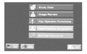
(3) Key Operator Functions—set up and manage system configurations (accessed by key operator and applications consultant only).
(4) Applications Consultant — change image processing parameters, access SMPTE Test Pattern.
(5) Service Functions — service the machine (qualified service personnel only).
2. Patient & exam data entry
Types of patient entries are as follows:
(1) New Patient一when information for a patient has never been entered in the CR system or the HIS/RIS system.
(2) Trauma一quick data entry fbr emergency conditions.
(3) Existing Patient一when information for the patient already exists in the CR system or the HIS/R1S system.
After entering the patient infbnnation, enter the exam information into the remaining fields on the Patient Input Screen. There are mandatory and optional fields as follows.
(1) Mandatory Exam Information: Cassette ID.
(2) Optional Exam Infbnnation: Body Part, Projection, Position, Orientation, Priority, Tech ID, Date of Birth, Gender, Procedure Name and Code, More Patient Information button, More Image Information button and Patient Location.
After you have entered the patient and exam information, touch submit. The information is stored in preparation for the exam.
3. Performing an exam
The procedure for performing an exam using a phosphor screen is the same as an exam using a screen/film.
To perform an exam, the procedure is as follows:
(1) Select the proper size cassette.
(2) Position the patient and the cassette.
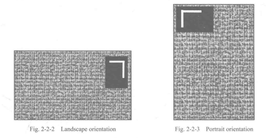
—For landscape orientation, place the yellow stripe label at the top right of the image when you position the patient, as is seen in Fig. 2-2-2.
一For portrait orientation, place the yellow stripe label at the top left of the image when you position the patient, as is seen in Fig. 2-2-3.
(3) Set the exposure factors.
(4) Expose the cassette.
(5) Place the cassette into the CR reader for scanning.
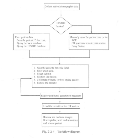
4. Working flow
The workflow of CR is seen in Fig. 2-2-4.
New Words
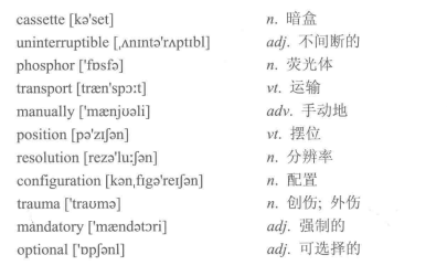
cassette [ko'set] | n.暗盒 |
uninterruptible [.AnmtsYAptibl] | adj.不间断的 |
phosphor ['fosfb] | n.荧光体 |
transport [treen'spait] | vt.运输 |
manually ['msenjuali] | adv.手动地 |
position [po'zijbn] | vt.摆位 |
resolution [rez0’lu:jbn] | n.分辨率 |
configuration [ksn.figaYeiJbn] | n.配置 |
trauma ['trauma] | n.创伤;外伤 |
mandatory ['m缶ndotori] | a#.强制的 |
optional [*DpJbnl] | adj.可选择的 |
Professional Phrases | |
1. bariumfluoride halide | 氟氯化裡 |
2. imaging plate | 成像板 |
3. latent image | 潜影 |
4. fluorescent light | 荧光 |
5. photo-multiplier tube | 光电倍增管 |
6. storage phosphor screen | 存储荧光屏 |
7. remote operational panel | 远程操作面板 |
8. Digital Imaging and Communications in Medicine (DICOM)医学数字图像及通信标准 | |
9. laser imager | 激光相机 |
10. uninterruptible power supply | 不间断电源 |
11. exposure factor | 曝光参数 |
12. trouble shooting | 故障排除 |
Key Notes to the Text
1. Conventional X-ray radiography uses film as imaging medium, realizes multi-functions of image acquisition, display, storage and transmission at one step, which limits the improvement of single function. Its biggest shortage is limited dynamic range.
传统的X射线摄影是以胶片作为成像介质,集图像釆集、显示、存储和传递多重功能 为一体,这样就限制了单个功能的改进。其最大的不足是动态范围有限。
2. After the exposure of X-ray, electrons of barium fluoride halide crystal, photo-active substance in IP, gain energy and jump to the higher energy level, leading to changes of crystafs structure.
经过X射线的照射后,成像板上的感光物质——氟氯化極晶体内的电子获得能量,跃 上较高的能级,引起晶体的结构发生变化。
3. The CR system operates in two basic modes: pass-through mode and quality assurance (QA) mode. The key operator, applications consultant, or service engineer configures the mode of operation under the direction of the department manager.
CR系统可在两种基本模式中运行,分别为流通模式和质量保证模式。主操作者、应用 顾问或者维修工程师可在部门经理的指导下配置相关的操作模式。
Item Detection
I ・ Answer the following questions according to the text.
1. Comparing with CR, what are the disadvantages of conventional X-ray radiography?
2. How can CR transform the information of human body into digital images?
3. How many modes does CR have? According to the text, who can configure the mode of operation?
4. To perform an exam successfully, which procedure should be paid attention to?
II. Translate the following professional words into Chinese.
1. analog2. laser3. storage4. modify
5. erase6. review7. gender8. database
HL Translate the following professional phrases into Chinese.
1. bar code scanner2. touch screen monitor3. imaging medium
4. dynamic range5. conventional film cassette6. local database
IV. Translate the following sentences into Chinese.
1. The CR system processes and produces digital images directly from latent images captured on imaging plate (IP).
2. Once the latent image is scanned, the electrons of IP release energy through fluorescent light and get back to its original level.
The fluorescent light is collected by photo-multiplier tube and is transformed into electrical signal.

