
Item 2 Digital X-ray Imaguig Equipment
Item Goals
Studying Objectives
In this item, digital X-ray imaging equipment are introduced, including computed radiography (CR), digital radiography (DR), digital subtraction angiography (DSA), digital gastrointestinal machine and laser imager. With the study of these items, it is essential to learn about the operating principle, operation procedure and systematic configuration of every equipment.
Knowledge Requirements
Mastery: To grasp some concepts, key points and professional terms about imaging process, operation procedure and systematic configuration of digital X-ray imaging equipment.
Acquaintance: To get acquainted with characteristics of CR, DR, DSA and digital gastrointestinal machine.
Understanding: To understand the components and operation of laser imager.
Section 1 Overview
Lead-in Questions
1. With the help of X-ray, have you ever thought of looking inside your body ? How can we transform the information of organs and tissues into digital images?
2. Could you list several kinds of digital X-ray imaging equipment?
3. In accordance with your knowledge, which characteristics should digital X-ray imaging equipment in the future have?
Text
X-ray is a form of electromagnetic wave. It behaves in the same way as light, but with
shorter wavelength.
When directed at a target of low density, X-ray can pass through the substance uninterrupted. Higher density targets will reflect or absorb X-ray. Thus, an X-ray image shows dark areas for soft tissues and shows light areas for bone. Passing through human body, it can carry important characteristic infbnnation of inner structure, in virtue of different density features among bone, tissue and muscle.
X-ray was discovered accidentally in 1895 by the German scientist Wilhelm Roentgen. Its most important application has been in medicine. X-ray revolutionizes the way how doctors detecte diseases and injuries. For the first time, we could see bones and other stnictures inside the living body instead of relying on symptoms, samples or surgery.
Since the first X-ray machine was invented, various kinds of X-ray machines have been developed for diagnosing and treating many diseases. In this item, we only discuss the diagnostic X-ray machine and focus on computed radiography (CR), digital radiography (DR), digital subtraction angiography (DSA) and digital gastrointestinal machine. Meanwhile, laser imager is also discussed for its application as auxiliary equipment.
Although there are great differences among diagnostic X-ray machines fbr different diagnostic objectives, the fundamental structure of X-ray machines is similar, which consists of X-ray generating device, X-ray imaging device and X-ray auxiliary device, as is shown in Fig. 2-1-1.
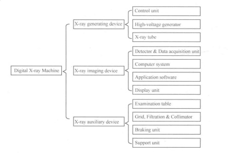
Fig. 2-1-1 System composition of X-ray machine
In X-ray generating device, X-ray tube is to generate X-ray, high-voltage generator is to supply filament voltage and tube voltage, and control unit takes charge of X-ray's quality, quantity and exposure time.
X-ray tube can transform electric energy into X-ray. High voltage generated by high- voltage generator is applied between cathode and anode of X-ray tube. It directly determines spectrum distribution of X-ray. Simultaneously, the controlled filament voltage acts upon the filament of X-ray tube and heats the cathode. Under a certain high voltage, the temperature of cathode determines the current of X-ray tube. Thus, the filament voltage determines the radiation quantity of X-ray.
X-ray auxiliary device contains kinds of mechanical devices to meet the needs of diagnosis. With the help of examination table and braking unit, it is convenient to position patient's area of interest into radiation field of X-ray tube. As to radiation field, collimator can adjust its rotary switch to change the range of radiation field in order to reduce unnecessary radiation. And grid can filter scattered radiation to improve image quality.
In the X-ray imaging device, detector and data acquisition unit of X-ray imaging device transform X-ray, passing through human body, into electrical signal. With the help of application software, display unit and computer, an image can be seen and post-processed by physicians.
The entire imaging process of X-ray machine can be seen in Fig. 2-1-2.
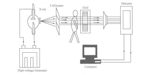
Fig. 2-1-2 Imaging process of X-ray machine
Based on the fundamental structure of X-ray machine, lots of medical departments have developed their own specialized X-ray machine, such as cardioangiography X-ray machine, whole-body angiography X-ray machine, mammogram X-ray machine, gastroenterography X-ray machine, X-ray machine for neurosurgery, and so on.
With the development of technology, the use of digital detector and the application of high-frequency high-voltage generator not only greatly improve the quality of X-ray image, but also decrease the radiation dose received by physicians and patients. However, there are new problems for digital medical imaging equipment, for example, how to realize effective management and make use of increasingly large digital information, how to mine more useful information from medical images, and so on. All the problems create the presence of RIS (radiology information system), PACS (picture archiving and communication system) and HIS (hospital information system).
New Words
electromagnetic [ijektrsm^g'netik] | adj.电磁的 |
wavelength [*weivleg0] | n.波长 |
generate ['djenoreit] | W.使形成;发生 |
auxiliary [Dig'zilisri] | n.辅助设备 |
grid [grid] | n.滤线栅 |
filtration [fiftrejon] | n.过滤板 |
collimator ['kolimeit^] | n.束线器 |
filament『fHomont] | n.灯丝 |
exposure [ik^pou^s] | n.曝光 |
cathode | n.阴极 |
radiation [reidfeijbn] | 〃.辐射 |
anode ['senaud] | n.阳极 |
transform [traens^rm] | vt.转换 |
application [.seplfkeijbn] | n.应用 |
detector [di'tekts] | n.探测器 |
substance [’sAbstons] | n.物质 |
radiography [.redi'ngrofi] | 放射线照相术;影像学 |
Professional Phrases
1. X-ray tube
2. digital X-ray machine
3. X-ray auxiliary device
4. high-voltage generator
5. data acquisition unit
6. application software
7. braking unit
8. filament voltage
9. tube voltage
10. exposure time
11. radiation quantity
12. post processing
13. computed radiography (CR)
14. digital radiography (DR)
15. digital substraction angiography (DSA)
16. digital gastrointestinal machine
17. radiography information system (RIS)
18. picture archiving and communication system (PACS)图片存档及通信系统
19. hospital information system (HIS)医院信息系统
Key Notes to the Text
1. When directed at a target of low density, X-ray can pass through the substance uninterrupted. Higher density targets will reflect or absorb the X-ray. Thus, an X-ray image shows dark areas for soft tissue and shows light areas for bone.
X射线能够穿透低密度物体。密度较高的目标则会反射或吸收X射线。因此,在X射 线图像中,软组织显示为喑区,而骨骼显示为亮区。
2. The controlled filament voltage acts upon the filament of X-ray tube and heats the cathode. Under a certain high voltage, the temperature of cathode determines the current of X-ray tube. Thus, the filament voltage determines the radiation quantity of X-ray.
受控的灯丝电压加于X射线管的灯丝,加热阴极。阴极温度决定了 X射线管的电流 (在一定高压下),因此灯丝电压决定了 X射线辐射量(管电流)O
3. As to radiation field, collimator, which is installed in the front of X-ray tube, can adjust its rotary switch to change the range of radiation field in order to reduce unnecessary radiation. And grid can filter scattered radiation to improve image quality.
提及辐射场,束线器通过调整旋转按钮改变辐射场的范围以减少不必要的辐射。滤线 栅则能够滤除散乱射线,以改善图像质量。
4. With the development of technology, there are new problems for digital medical imaging equipment, for example, how to realize effective management and make use of increasingly large digital information, how to mine more useful information from these kinds of information, and so on.
随着科技的发展,数字化医学影像设备出现了新的问题,例如,如何对日益庞大的数 字化信息进行有效的管理及利用,以及如何从医学图像中挖掘更多的有用信息等。
Item Detection
I ・ Answer the following questions according to the text.
1. Why does the image show dark areas for soft tissue and light areas for bone?
2. How many equipment are discussed in this item? Please list these equipment.
3. What is the fundamental structure of X-ray machines? And could you list the components of every part?
4. How can X-ray machine control the quantity and quality of X-ray?
5. In accordance with the development of technology, could you imagine the X-ray machine in the future?
II. Translate the following professional words into Chinese.
1. digital2. quality3. quantity4. angiography5. gastrointestinal
6. spectrum7. density8. characteristic9. symptom
III. Translate the following professional phrases into Chinese.
1. control unit2. X-ray imaging device3. X-ray generating device
4. display unit5. spectrum distribution6. mechanical device
7. specialized X-ray machine
IV. Translate the following sentences into Chinese.
1. Passing through human body, it can carry important characteristic infbmiation of inner structure, due to different density features among bone, tissue and muscle.
2. Although there are great differences among diagnostic X-ray machines for different diagnostic objectives, the fundamental structure of X-ray machines is similar, which consists of X-ray generating device, X-ray imaging device and X-ray auxiliary device.
3. In the X-ray generating device, X-ray tube is to generate X-ray, high-voltage generator is to supply filament voltage and tube voltage, and control unit takes charge of X・ray's quality, quantity and exposure time.
4. In the X-ray imaging device, detector and data acquisition unit of X-ray imaging device transform X-ray, passing through human body, into electrical signal.

